Radiology
During our comprehensive examination in our clinics; You will be informed about your general health status and, if any, medications you use regularly; Your general oral examination is performed, your expectations from the treatment are evaluated and an x-ray examination for diagnostic purposes may be requested.
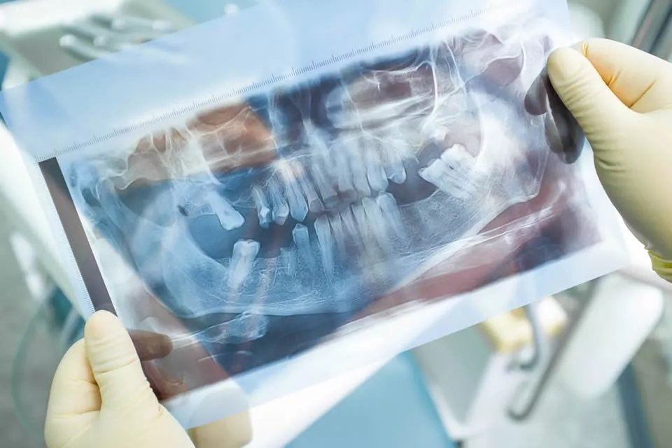
During your general oral examination, all your teeth and intraoral tissues will be evaluated and you will be informed if any interventions related to teeth that you do not have any complaints about yet. Tooth decays that are not deep yet may not bother you, but you may feel discomfort when it progresses. Or, if you are informed about your gingival disease in the initial stage, you can prevent future gingival recessions and tooth loss. After your oral examination, various x-ray examinations may be requested for diagnostic purposes. Generally, it may be necessary to take a Panoramic X-ray in cases where the evaluation of the entire intraoral condition is required, or in cases of complaints caused by a single tooth locally, the condition of the relevant teeth can be evaluated by taking intraoral X-rays. Since the panoramic x-ray gives a general view of the jaws, it sometimes enables the diagnosis of conditions such as interface caries, incompatible restoration, incomplete canal treatment, root tip inflammation that cannot be detected during oral examination.
Today, it is an indispensable diagnostic method for medical people. It is an auxiliary diagnosis method.
It is the beams that contribute to the diagnosis without opening the tissues of the patient, touching the patient, touching the patient without disturbing him.
When exposed to traumas, fractures, the size and position of pathologies, impacted teeth, teeth. They are useful in evaluating their position in the jawbone.
It is an indispensable method we apply in cases where we cannot evaluate the oral cavity clinically, and when we need to see inside the jaw bones.
No its not. The dose of radiation from a single dental x-ray film is even less than the radiation received when exposed to sunlight. There is no medical harm in taking 12-14 dental x-rays from the whole mouth at one time.
- For diagnosis and diagnosis
- During treatments,
- For control after treatments
- For follow-up purposes to monitor lesions in certain periods < / ul>
The risk of initiation of LEUKEMIA is directly related to the amount and dose of irradiated blood-producing tissues. In dental radiography applications, the irradiated areas of the maxilla and mandible contain a very small part of the active bone marrow. As a result, approximately 2000-5000 periapical films should be taken for leukemia depending on the periapical films taken from the patient.
It is one of the tissues with the highest rate of cancer caused by radiation. Although the thyroid gland is not irradiated with the primary beam during dental radiographic procedures, thyroid radiation exposure occurs. In dental radiology, there are serious doubts about the radiation that the thyroid gland receives because it is close to the x-ray beam. Thyroid dose is not as high as thought in dental radiology applications. The amount of radiation taken in panoramic radiography is 1% of the thyroid dose resulting from cervical spinal examinations. In dental radiology, the patient should be dressed in a lead collar with a lead apron to protect the thyroid gland.
When the patient is put on a standard 0.5 mm thick lead apron, the amount of radiation the patient receives decreases by 95%.
Unless necessary, x-rays for dental problems should be avoided. If necessary, it should be pulled by wearing lead apron.
It is very important to use thyroid protectors, but it is not appropriate to use a lead collar during the shooting of the cephalometric films taken for orthodontic purposes and panoramic radiographs that are in routine use.
In order to protect from radiation damage, pregnant or pregnancy possibility patients should definitely warn their physicians and radiation officers. In addition, companions are also prohibited from entering the X-ray Clinic.
Panoramic Radiography: It is a film shooting technique in which the upper and lower tooth curves and adjacent tissues and formations are displayed on a single film. used to detect. It can detect problematic teeth and help diagnose cysts and tumors.
Gives dentists general information about teeth and jaws in treatment planning, with the general appearance and details it gives.
Periapical Radiography: It is a type of small film that gives the closest view of the teeth to the exact size.
It gives a detailed image of the related tooth group and the jawbone around it.
Bite-Wing Radiography (Biting radiography): It is especially used to detect caries between teeth and neighboring teeth.
Hand-Wrist Radiography: It is used to determine the bone age of pediatric patients in orthodontic treatment planning.
Cephalometric Radiography: It is especially used in orthodontic treatment planning, the two-dimensional relationship of the lower and upper jaws and teeth with the base of the skull and other tissues is evaluated.
TMJ Radiography: Temporomandibular joint (jaw joint) moire
Occlusal Radiography: It is used to determine the horizontal positions of the teeth, to examine the formations in the jawbone or to examine the salivary gland diseases.

